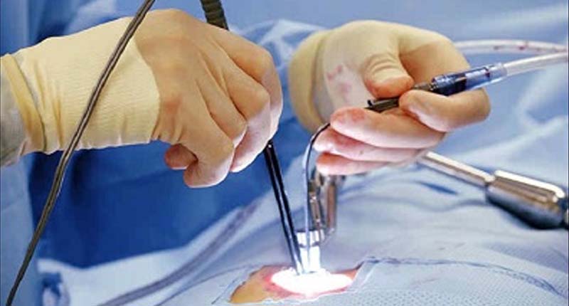
With these smaller incisions, there's less trauma or disruption of the soft tissue around the spine, which typically results in less blood loss lower rates of infection and faster recovery.
The indications for minimally invasive spine surgery are relatively similar to open surgery. Several conditions can be treated with this technique, such as:
- Stenosis which is narrowing around the spinal cord or nerve roots.
- Infections of the spine.
- Tumor.
- Diminished sensation to heat & cold.
- Instability in the spine which is the abnormal movement between bones which can be very painful. You will find the Best Specialist for Minimally Invasive Surgery in Kandivali, Mumbai is Dr. Yash Shah, Provides highly advanced treatment. Book Appointment Today !
Advantage of Minimally Invasive Spine Surgery
- Stenosis which is narrowing around the spinal cord or nerve roots.
- Smaller incisions, so there's less disruption of the soft tissue around the spine patients.
- Less pain after surgery.
- Lower rates of infection.
- Patients can recover and get back to life quicker.
In general, there are two types of minimally invasive surgery.
1. Decompression
- In this minimally invasive surgery, we remove bones or ligaments strategically, and in some cases, disk fragments.
- compressing the spinal cord or nerve roots.
- A typical retractor is inserted down through the muscle and split the muscle fibers, and then we're able to work with long curved instruments through the retractor to accomplish the work.
2. Spine Fusion Surgery
In Spine Fusion minimally invasive spine surgery, surgeons try to decompress the nerves and spinal cord but also fusing or fixing the vertebrae together. This eliminates painful movement between the bones. With spinal fusion surgery, we typically have to place hardware into the spine, such as plates screws or rods, to eliminate the movement.
In the lumbar spine, we remove the disk space, which is a gelatinous shock absorber between the bones, and after we remove the disk, we typically place a spacer in the disk space. This allows the bone to grow through the spacer and around the spacer together so that this part of the vertebra becomes a single bone.
In the lumbar spine, we remove the disk space, which is a gelatinous shock absorber between the bones, and after we remove the disk, we typically place a spacer in the disk space. This allows the bone to grow through the spacer and around the spacer together so that this part of the vertebra becomes a single bone.


